Loading...
Categories

Thematic groups

Funding periods

Project type

Top keywords
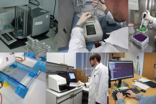
Picture: The collage contains six photographs of steps in molecular analyses in a laboratory, performed by a scientist. Photo 1 shows a sample standardization device. Photo 2 shows the scientist working on a polymerase chain reaction device. Photo 3 shows the scientist using an electronic pipette to fill a sample into a micro-reaction tube. Photo 4 shows an Illumina MiSeq sequencer. Photo 5 shows the scientist working on a bioinformatics quality control device. Photo 6 shows the scientist at a desk in front of a computer making inputs for assignment.
Microorganisms (Core project)
#Microorganisms & Fungi  #BEF  #Soil Ecology  #REX/LUX  #FOX  #2023 – 2026  #2020 – 2023  #Fungi […]
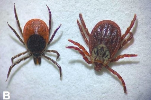
Picture: The photo shows a close-up of two female ticks, on the left a specimen of Ixodes ricinus and on the right a specimen of Dermacentor reticulatus.
LaBiRo (Contributing project)
#Animals  #2023 – 2026  #2020 – 2023  #Species biodiversity […]
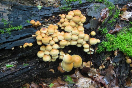
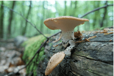
Figure: The photo shows a group of light brown mushrooms growing on a deadwood tree trunk.
WOODSTOCK I (Contributing project)
#Forest & Deadwood  #BELongDead  #2020 – 2023  #Ecosystem services […]
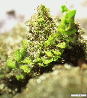
Picture: The photo shows a close-up of liverwort.
Crustfunction III (Contributing project)
#Soil biology & Element cycling  #Soil Ecology  #FOX  #2023 – 2026  #2020 – 2023  #Phosphorus cycle […]
Picture: The photo shows a mushroom growing on a deadwood tree trunk lying in the forest
BLD-MFD-HZG III (Contributing project)
#Forest & Deadwood  #BELongDead  #FOX  #2020 – 2023  #Deadwood […]
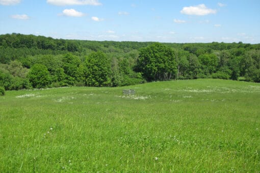
Figure: The photo shows a hilly landscape with a meadow and a deciduous forest under a blue sky in spring.
BE_CH4 (Contributing project)
#Microorganisms & Fungi  #2017 – 2020  #Forest […]
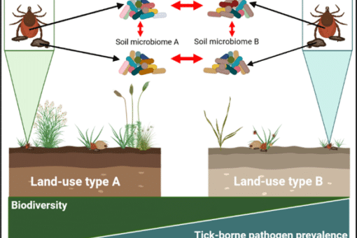
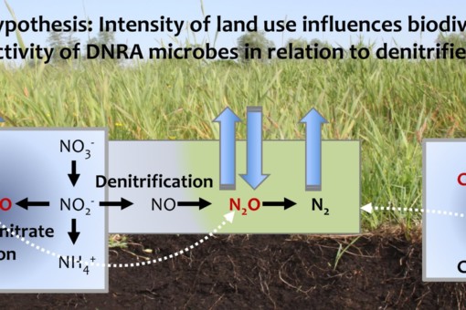
Picture: The graphic shows information on the central thesis of the project.
BE-Cult (Contributing project)
#Microorganisms & Fungi  #2017 – 2020  #Nitrogen cycle […]
Picture: The photo shows a close-up of liverwort.
Crustfunction II (Contributing project)
#Soil biology & Element cycling  #2017 – 2020  #Species communities […]
Picture: The collage contains six photographs of steps in molecular analyses in a laboratory, performed by a scientist. Photo 1 shows a sample standardization device. Photo 2 shows the scientist working on a polymerase chain reaction device. Photo 3 shows the scientist using an electronic pipette to fill a sample into a micro-reaction tube. Photo 4 shows an Illumina MiSeq sequencer. Photo 5 shows the scientist working on a bioinformatics quality control device. Photo 6 shows the scientist at a desk in front of a computer making inputs for assignment.
Microorganisms (Core project)
#Microorganisms & Fungi  #2017 – 2020  #Species communities […]
Picture: The collage contains six photographs of steps in molecular analyses in a laboratory, performed by a scientist. Photo 1 shows a sample standardization device. Photo 2 shows the scientist working on a polymerase chain reaction device. Photo 3 shows the scientist using an electronic pipette to fill a sample into a micro-reaction tube. Photo 4 shows an Illumina MiSeq sequencer. Photo 5 shows the scientist working on a bioinformatics quality control device. Photo 6 shows the scientist at a desk in front of a computer making inputs for assignment.
Microorganisms (Core project)
#Microorganisms & Fungi  #2014 – 2017  #Species communities […]
Picture: The photo shows a close-up of liverwort.
Crustfunction I (Contributing project)
#Soil biology & Element cycling  #2014 – 2017  #Species communities […]
Top