The projects of the
Biodiversity Exploratories
Currently, there are more than 40 projects involved. They investigate the cause-and-effect relations in the enlivened nature, both, by monitoring as well as experiments. Use the filter options to search for specific projects.
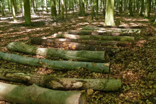
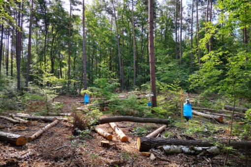
BELongDead (Contributing project)
#Forest & Deadwood #BELongDead #2023 – 2026 #2020 – 2023 #2017 – 2020 #2014 – 2017 #2011 – 2014 #2008 – 2011
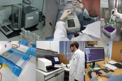
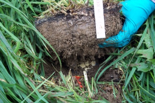
Microorganisms (Core project)
#Microorganisms & Fungi #BEF #Soil Ecology #REX/LUX #FOX #2020 – 2023
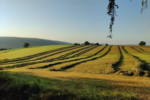
MultiCrossBEF (Cooperation project)
#Animals #Soil biology & Element cycling #Plants #Ecosystem function of biodiversity #Biotic Interaction #2023 – 2026
Microorganisms (Core project)
#Microorganisms & Fungi #BEF #Soil Ecology #REX/LUX #FOX #2023 – 2026
BEMPL (Contributing project)
#Microorganisms & Fungi #2023 – 2026 #2020 – 2023
EXClAvE I (Contributing project)
#Plants #BEF #2020 – 2023
SeedDiv (Contributing project)
#Plants #Plants #BEF #2023 – 2026 #2020 – 2023
SMaTI (Contributing project)
#Animals #BEF #Biotic Interaction #REX/LUX #2023 – 2026 #2020 – 2023
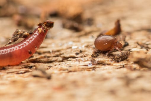
SPIDERFUN (Contributing project)
#Animals #BEF #Biotic Interaction #Soil Ecology #2023 – 2026 #2020 – 2023
SPRINT (Contributing project)
#Animals #Biotic Interaction #REX/LUX #2020 – 2023
TrophCost (Contributing project)
#Theory, Modelling & Upscaling #Biotic Interaction #Social-Ecology #2023 – 2026 #2020 – 2023
Crustfunction III (Contributing project)
#Soil biology & Element cycling #FOX #Soil Ecology #2023 – 2026 #2020 – 2023
GapResin (Contributing project)
#Animals #FOX #2020 – 2023
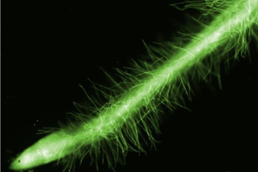
HAIRphae (Contributing project)
#Plants #BEF #Remote Sensing, Lanscape and Modelling #2023 – 2026 #2020 – 2023
SCALEMIC (Contributing project)
#Microorganisms & Fungi #BEF #Soil Ecology #2023 – 2026 #2020 – 2023
LitterLinks (Contributing project)
#Animals #Soil biology & Element cycling #2020 – 2023
HiLUCC (Contributing project)
#Theory, Modelling & Upscaling #Remote Sensing, Lanscape and Modelling #2020 – 2023 #2017 – 2020
FuncNet (Contributing project)
#Animals #BELongDead #2020 – 2023 #2017 – 2020
RESOILIENCE (Contributing project)
#Animals #BEF #Soil Ecology #2020 – 2023 #2017 – 2020
SADE (Contributing project)
#Plants #2020 – 2023 #2017 – 2020 #2014 – 2017
MicroBEEs (Contributing project)
#Animals #2017 – 2020
SCALEMIC (Contributing project)
#Microorganisms & Fungi #2017 – 2020
Crustfunction II (Contributing project)
#Soil biology & Element cycling #2017 – 2020
Microorganisms (Core project)
#Microorganisms & Fungi #2017 – 2020
HEDGE II (Contributing project)
#Theory, Modelling & Upscaling #2017 – 2020
MetacommuniTree (Contributing project)
#Animals #2014 – 2017
SCALEMIC (Contributing project)
#Microorganisms & Fungi #2014 – 2017
Microorganisms (Core project)
#Microorganisms & Fungi #2014 – 2017
LitterLinks (Contributing project)
#Animals #Soil biology & Element cycling #2014 – 2017
HEDGE I (Contributing project)
#Theory, Modelling & Upscaling #2014 – 2017
SCALEMIC (Contributing project)
#Microorganisms & Fungi #2011 – 2014
LitterLinks (Contributing project)
#Animals #Soil biology & Element cycling #2011 – 2014 #2008 – 2011
LitterLinks (Contributing project)
#Animals #Soil biology & Element cycling #2017 – 2020
SCALEMIC (Contributing project)
#Microorganisms & Fungi #2008 – 2011
Crustfunction I (Contributing project)
#Soil biology & Element cycling #2014 – 2017