Loading...
Categories

Thematic groups

Funding periods

Project type

Top keywords
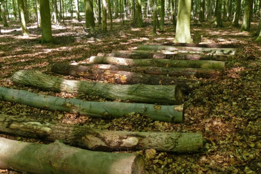
Figure: The photo shows a row of deadwood logs in a shady summer forest.
BELongDead (Contributing project)
#Forest & Deadwood  #BELongDead  #2023 – 2026  #2020 – 2023  #2017 – 2020  #2014 – 2017  #2011 – 2014  #2008 – 2011  #Fungi […]
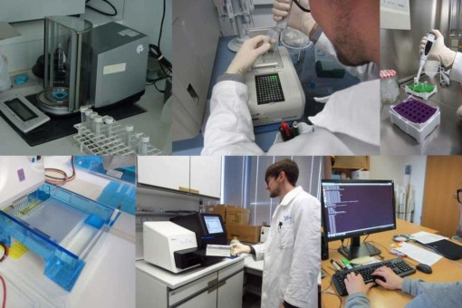
Picture: The collage contains six photographs of steps in molecular analyses in a laboratory, performed by a scientist. Photo 1 shows a sample standardization device. Photo 2 shows the scientist working on a polymerase chain reaction device. Photo 3 shows the scientist using an electronic pipette to fill a sample into a micro-reaction tube. Photo 4 shows an Illumina MiSeq sequencer. Photo 5 shows the scientist working on a bioinformatics quality control device. Photo 6 shows the scientist at a desk in front of a computer making inputs for assignment.
Microorganisms (Core project)
#Microorganisms & Fungi  #BEF  #Soil Ecology  #REX/LUX  #FOX  #2020 – 2023  #Nitrogen cycle […]
Picture: The collage contains six photographs of steps in molecular analyses in a laboratory, performed by a scientist. Photo 1 shows a sample standardization device. Photo 2 shows the scientist working on a polymerase chain reaction device. Photo 3 shows the scientist using an electronic pipette to fill a sample into a micro-reaction tube. Photo 4 shows an Illumina MiSeq sequencer. Photo 5 shows the scientist working on a bioinformatics quality control device. Photo 6 shows the scientist at a desk in front of a computer making inputs for assignment.
Microorganisms (Core project)
#Microorganisms & Fungi  #BEF  #Soil Ecology  #REX/LUX  #FOX  #2023 – 2026  #Nitrogen cycle […]
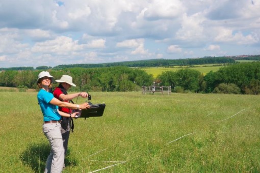
Picture: The photo shows an unmown meadow with an area marked with white tape. In the foreground are project leader Professor Anja Linstädter and PhD student Florian Männer, both wearing hats to protect them from the sun. Ms. Linstädter is holding a black flat box in her hands at waist level. Inside the box is a white reference plate over which Mr. Männer is holding a field spectrometer to calibrate it. In the background of the image is a fenced climate measurement station and a row of bushes and trees. Meadows and forests can be seen on the horizon
SeBAS (Contributing project)
#Theory, Modelling & Upscaling  #BEF  #Plants  #Remote Sensing, Lanscape and Modelling  #Social-Ecology  #2023 – 2026  #2020 – 2023  #Spatial scale […]
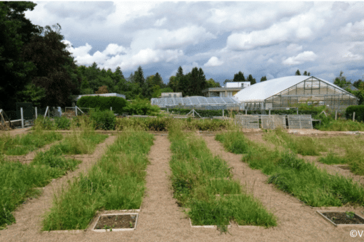
Picture: The photo shows the experimental garden of the Philipps University in Marburg, Germany, called "Common Garden". You can see swards with tall growing grasses in six rows of square sub-plots of fifty by fifty centimeters, where always two sub-plots are laid out next to each other. One row is thus one meter wide. Between the rows are paths. In the background are greenhouses, buildings and behind them a dense row of deciduous and coniferous trees under a sky covered with cumulus clouds
EXClAvE I (Contributing project)
#Plants  #BEF  #2020 – 2023  #Cultivation […]
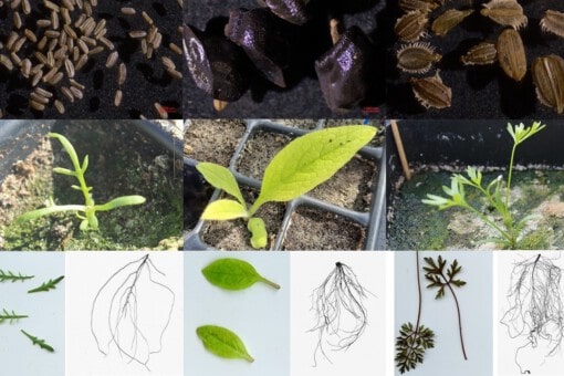
Picture: The collage shows twelve photos of different seeds and seedlings.
SeedDiv (Contributing project)
#Plants  #BEF  #Plants  #2023 – 2026  #2020 – 2023  #Species communities […]
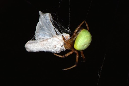
Picture: The photo shows a pumpkin spider, Latin Araniella cucurbitina, spinning a victim in front of a black background. The round abdomen of the spider is bright light green, while the rest of the body is brown.
SPIDERFUN (Contributing project)
#Animals  #BEF  #Biotic Interaction  #Soil Ecology  #2023 – 2026  #2020 – 2023  #Traits […]
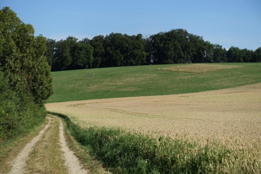
Picture: The photo shows a slightly hilly landscape in summer under a blue sky. In the front left of the picture there are high bushes, a tree and a field path that turns to the left further back. From the middle to the right to the edge of the picture there is a field with beige-coloured ears of grain. Behind the field there is a mown meadow with short green grass on a slightly rising hill. On the left of the meadow there is a fenced climate measuring station. On the right of the meadow there is an unmown experimental area with tall brown and green grass. Behind the meadow there is a dense row of deciduous trees running through the picture.
SPRINT (Contributing project)
#Animals  #Biotic Interaction  #REX/LUX  #2020 – 2023  #Grassland […]
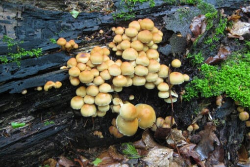
Figure: The photo shows a group of light brown mushrooms growing on a deadwood tree trunk.
WOODSTOCK I (Contributing project)
#Forest & Deadwood  #BELongDead  #2020 – 2023  #Nitrogen cycle […]
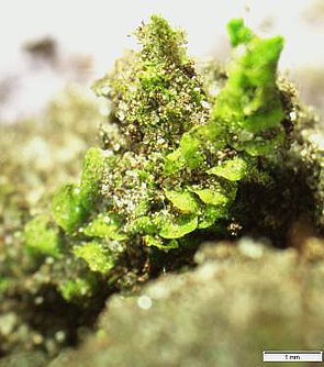
Picture: The photo shows a close-up of liverwort.
Crustfunction III (Contributing project)
#Soil biology & Element cycling  #Soil Ecology  #FOX  #2023 – 2026  #2020 – 2023  #Soil hydrology […]
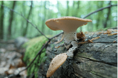
Picture: The photo shows a mushroom growing on a deadwood tree trunk lying in the forest
BLD-MFD-HZG III (Contributing project)
#Forest & Deadwood  #FOX  #BELongDead  #2020 – 2023  #Fungi […]
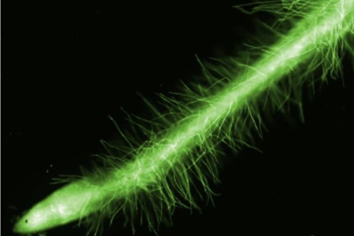
Picture: The macroscopic photo shows the hairs of a root in bright green color against a black background
HAIRphae (Contributing project)
#Plants  #BEF  #Remote Sensing, Lanscape and Modelling  #2023 – 2026  #2020 – 2023  #Soil […]
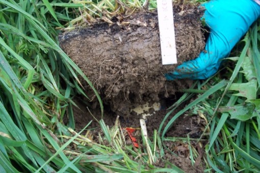
Picture: The photo shows a hand in a turquoise glove lifting a piece of soil between meadow grass. The thickness of the piece of soil is estimated at ten to fifteen centimetres.
SCALEMIC (Contributing project)
#Microorganisms & Fungi  #BEF  #Soil Ecology  #2023 – 2026  #2020 – 2023  #Nitrogen cycle […]
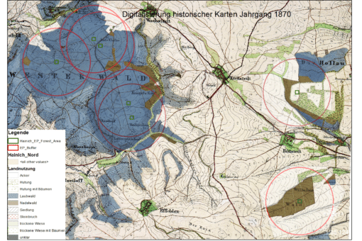
Picture: The drawing shows a historical map of the Hainich from the year eighteen hundred and seventy.
HiLUCC (Contributing project)
#Theory, Modelling & Upscaling  #Remote Sensing, Lanscape and Modelling  #2020 – 2023  #2017 – 2020  #Land use […]
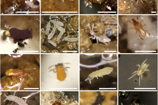
Picture: The collage includes sixteen photographs of soil-dwelling Acari and Collembola taken from a micro-arthropod sample from the Biodiversity Exploratory Greenlands.
RESOILIENCE (Contributing project)
#Animals  #BEF  #Soil Ecology  #2020 – 2023  #2017 – 2020  #Microbial community […]
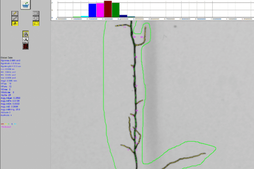
Picture: The screenshot shows the image analysis of a root system with the program Winrhizo
RootFun (Contributing project)
#Plants  #2020 – 2023  #2017 – 2020  #Mycorrhiza fungi […]
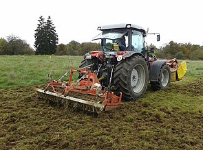
Picture: The photo shows a tractor with trailed implement tearing up the meadow surface to cause a disturbance.
SADE (Contributing project)
#Plants  #2020 – 2023  #2017 – 2020  #2014 – 2017  #Nutrients […]
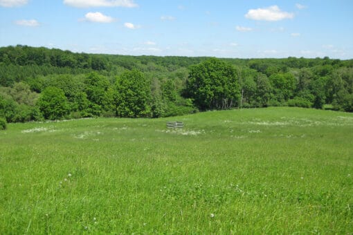
Figure: The photo shows a hilly landscape with a meadow and a deciduous forest under a blue sky in spring.
BE_CH4 (Contributing project)
#Microorganisms & Fungi  #2017 – 2020  #Microbial community […]
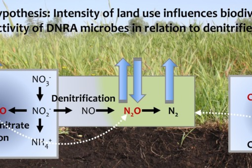
Picture: The graphic shows information on the central thesis of the project.
BE-Cult (Contributing project)
#Microorganisms & Fungi  #2017 – 2020  #Climate change […]
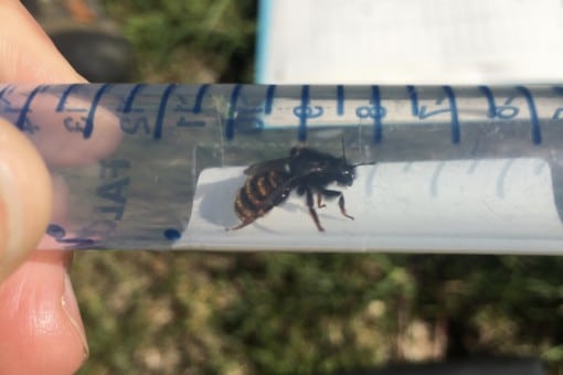
Picture: The photo shows a captured, living mason bee in a transparent plastic tube with blue printed graduations, which is held horizontally in front of the camera. In the background meadow soil can be seen.
MicroBEEs (Contributing project)
#Animals  #2017 – 2020  #Ecosystem processes […]
Picture: The photo shows a close-up of liverwort.
Crustfunction II (Contributing project)
#Soil biology & Element cycling  #2017 – 2020  #Mineralisation […]
Picture: The collage contains six photographs of steps in molecular analyses in a laboratory, performed by a scientist. Photo 1 shows a sample standardization device. Photo 2 shows the scientist working on a polymerase chain reaction device. Photo 3 shows the scientist using an electronic pipette to fill a sample into a micro-reaction tube. Photo 4 shows an Illumina MiSeq sequencer. Photo 5 shows the scientist working on a bioinformatics quality control device. Photo 6 shows the scientist at a desk in front of a computer making inputs for assignment.
Microorganisms (Core project)
#Microorganisms & Fungi  #2017 – 2020  #Nitrogen cycle […]
Picture: The photo shows a hand in a turquoise glove lifting a piece of soil between meadow grass. The thickness of the piece of soil is estimated at ten to fifteen centimetres.
SCALEMIC (Contributing project)
#Microorganisms & Fungi  #2014 – 2017  #Soil […]
Picture: The collage contains six photographs of steps in molecular analyses in a laboratory, performed by a scientist. Photo 1 shows a sample standardization device. Photo 2 shows the scientist working on a polymerase chain reaction device. Photo 3 shows the scientist using an electronic pipette to fill a sample into a micro-reaction tube. Photo 4 shows an Illumina MiSeq sequencer. Photo 5 shows the scientist working on a bioinformatics quality control device. Photo 6 shows the scientist at a desk in front of a computer making inputs for assignment.
Microorganisms (Core project)
#Microorganisms & Fungi  #2014 – 2017  #Nitrogen cycle […]
Picture: The photo shows a hand in a turquoise glove lifting a piece of soil between meadow grass. The thickness of the piece of soil is estimated at ten to fifteen centimetres.
SCALEMIC (Contributing project)
#Microorganisms & Fungi  #2011 – 2014  #Soil […]
Picture: The photo shows a hand in a turquoise glove lifting a piece of soil between meadow grass. The thickness of the piece of soil is estimated at ten to fifteen centimetres.
SCALEMIC (Contributing project)
#Microorganisms & Fungi  #2008 – 2011  #Soil […]
Picture: The photo shows a close-up of liverwort.
Crustfunction I (Contributing project)
#Soil biology & Element cycling  #2014 – 2017  #Mineralisation […]
Top